


Nonspecific T Wave Abnormality
Having T wave abnormality, nonspecific, on an EKG or ECG report can be quite upsetting if you’re not sure what the phrase means.
The key point is that nonspecific T wave abnormality is simply a description of an ECG pattern; it is not a diagnosis. Your T wave is just one of the components of your body’s electrical pattern that is picked up and translated into an ECG. Because it simply reflects how the cells of your heart are functioning, many factors can affect it and produce a nonspecific pattern. Different people can have the same general pattern with different underlying conditions and this makes the T wave changes seem confusing when seen by themselves.
Therefore if your question is ‘Nonspecific T wave abnormality- is it dangerous?’ The answer would be always depends. What does it really mean is that it will vary greatly based on its pattern, what leads are involved, other ECG findings, if you are symptomatic or not, if you are otherwise healthy or not, and the timing of its appearance.
What Does the T Wave Represent on an ECG?
An electrocardiogram is a test used to record the heart’s electrical activity.The T wave is indicative of the process of ventricular repolarization, which is when the ventricles recover electrically after having been activated to pump blood. The American Heart Association, American College of Cardiology Foundation and Heart Rhythm Society define the T wave as representing the rapid repolarization phase of the ventricles.
There’s no need to understand the cellular biology involved to understand the statement on a report.
A simplified ECG contains several recognizable components:
P wave → QRS complex → T wave
T wave T wave shape The appearance of the T wave depends on the lead. T wave shape also depends on age among other factors; hence, the T wave shape should not look the same for all 12 leads.
Why does this matter? It can look very much like a T wave change when it appears in one lead but we do not automatically see it as the same change if it appears across multiple leads.
What Does Nonspecific T Wave Abnormality Mean?
A nonspecific T wave abnormality means the T-wave appearance is outside an expected pattern, but the finding does not point clearly to one specific cause.
The word “nonspecific” is the key.
It does not mean:
- “There is definitely nothing wrong.”
- “You have heart disease.”
- “You are having a heart attack.”
- “The ECG is completely normal.”
But it means that the mere finding of the ECG is too nondescript to be a specific explanation.
AHA/ACCF/HRS guidelines on T-wave abnormalities mention that simple T-wave abnormalities are heterogenous on their etiology and that comparative evaluation of a previous ECG is important.
So a clinician has to look at the overall picture of the ECG and the person to arrive at a conclusion rather than focusing on a sentence in the report alone.
What Can a Nonspecific T Wave Abnormality Look Like?
A nonspecific T wave abnormality can involve changes such as T-wave flattening, low amplitude, or mild inversion, depending on the ECG pattern and lead involved.
T-Wave Flattening
A flattened T wave has less height than expected.
The American Heart Association’s ECG standardization recommendations use descriptive terms such as “flat,” “low,” and “inverted” for T-wave morphology.
T-Wave Inversion
When the T wave is going in the opposite direction to what you normally see in a specific lead, this is T-wave inversion.
But not all T-wave inversion is the same. Certain T-wave formations can be normal by lead, age and other factors.
Biphasic T Waves
A biphasic T wave has both positive and negative portions. This is another descriptive ECG feature that requires interpretation in context.
Nonspecific ST-T Abnormality
Sometimes a report refers to a nonspecific ST-T abnormality rather than only the T wave. This indicates that nonspecific changes involve the ST segment, T wave, or both.
Quick comparison
| ECG finding | What it describes |
| Nonspecific T wave abnormality | T-wave appearance that does not identify one specific cause |
| T-wave flattening | Reduced T-wave amplitude |
| T-wave inversion | T wave points downward in a lead where that pattern may be abnormal |
| Biphasic T wave | T wave has positive and negative components |
| Nonspecific ST-T abnormality | Nonspecific changes involving the ST segment and/or T wave |
These descriptions should not be used to diagnose yourself from an ECG image. Lead location, the rest of the tracing, symptoms, and clinical history all matter.
Nonspecific T Wave Abnormality: Is It Dangerous?
If you searched “nonspecific T wave abnormality is it dangerous,” the best you can do is: because it is a words alone, you can’t diagnose the severity of it.
Some nonspecific T-wave changes, however, can be present without an immediately severe pathology. T-wave abnormalities are also present in patients with cardiovascular or other systemic illnesses.
The distinction is important.
A 2026 study in Heart revealed isolated nonspecific ST-T abnormalities were linked to higher heart failure risk over a follow-up period. However, this is an observational association, not proof that a person with a nonspecific T-wave abnormality has or will develop heart failure.
Because of that new evidence, it is also better to steer clear of labeling every single nonspecific ECG abnormality as 100% safe or simply unsafe.
Instead, think of it as a finding that needs appropriate context.
Factors That Can Change Its Significance
A clinician may consider:
- Whether you have symptoms
- Whether the ECG finding is new
- Whether a previous ECG showed the same pattern
- Which ECG leads are affected
- Whether other ECG abnormalities are present
- Your medical history
- Relevant cardiovascular risk factors
- Medications
- Relevant laboratory findings
The American Heart Association also explains that ECG patterns can occur in different structural and physiologic states, which limits the ability of one ECG pattern to identify a single cause and highlights the importance of understanding electrocardiogram (ECG) results in context.
What Can Cause Nonspecific T Wave Abnormalities?
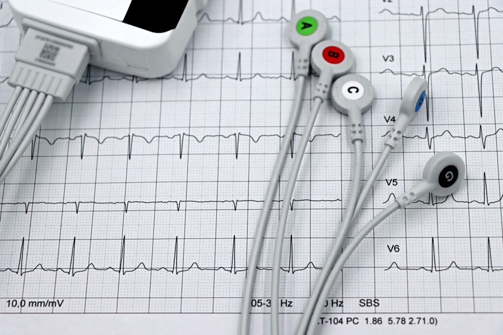
There is no single cause.
Possible contributors include cardiac conditions, electrolyte disturbances, medications, changes in ventricular activation, physiologic factors and technical factors affecting the ECG.
| Category | Examples or contributors |
| Cardiac factors | Ischemia, ventricular hypertrophy, cardiomyopathy and other cardiac conditions |
| Electrolyte/metabolic factors | Abnormal potassium and other electrolyte levels |
| Medications | Some medicines can alter cardiac repolarization |
| Physiologic factors | Changes in heart rate, breathing and other physiologic states |
| Conduction/activation changes | Bundle branch block, ventricular pacing and other altered ventricular activation |
| Technical factors | Electrode placement or recording-related issues |
| Normal variation | Some T-wave patterns vary with age and ECG lead |
As explained in AHA/ACCF/HRS scientific statement, primary repolarization abnormalities can be related to ischemia, myocarditis, drugs, toxins and electrolytes abnormalities; physiologic alterations like heart rate, hyperventilation, body position and temperature can affect repolarization. When ventricular depolarization is changed secondary ST-T changes can also happen.
This is the reason you need not assume that a comprehensive list of what could be wrong with you is also a list of what you can diagnose yourself.
Nonspecific T Wave Abnormality vs. T Wave Inversion
These terms are related but are not interchangeable.
A nonspecific T wave abnormality is a broader description. T-wave inversion describes a particular direction of the T wave.
For instance, an ECG report may be recording of T-wave abnormality of various incomplete forms without specifying a particular illness. On other hand, the judge may specify a more obvious inversion pattern that should be given a diffrerent assess.
AHA/ACCF/HRS recommendations: use of descriptive terminology Justification for the guidelines: Isolated T-wave abnormalities can cause confusion or false positive interpretation.
The importance of this distinction is that one cannot assume that the notation of “T-wave abnormality” implies the same as an identifiable pathologic T-wave inversion pattern.
Why ECG Lead Location Matters
What an ECG Doesn’t Show. An ECG doesn’t show the heart from a single perspective.
The 12-lead ECG records electrical activity from different angles. The shape of a T wave in a normal 12-lead ECG is not the same in each lead and there are certain normal shapes of T wave in normal patients that are age dependent.
For instance, AHA/ACCF/HRS guidelines specify the expected degree of T-wave variation between leads, between age groups, etc.
This means that interpreting a T wave requires more than asking:
“Is the T wave upright or inverted?”
A clinician also considers where the change appears and whether the pattern fits the rest of the ECG, along with established guidance for electrocardiogram interpretation.
That is another reason a single phrase from an automated ECG report cannot provide a complete diagnosis.
Why Previous ECGs Are Important
One of the most useful pieces of information can be an older ECG.
Suppose a current report contains a nonspecific T wave abnormality. If an ECG from several years earlier contains essentially the same finding, that provides different context from a brand-new change.
Comparison can help answer questions such as:
- Is this finding new?
- Has it been present for a long time?
- Has the pattern changed?
- Are there additional ECG changes now?
Comparison to prior ECGs is specifically mentioned in AHA/ACCF/HRS guidancen in the context of evaluating mild T-wave abnormalities.
Nor does it mean that an old lesion can be automatically discarded. It does, however, offer an additional finding to interpret clinically.
Why Does an ECG Report Say “Nonspecific”?
Computer analysis programs are commonly used in today’s ECG systems for measurement and pattern analysis.
Hence the wording on an ECG report can be a statement that is a generic interpretive remark rather than the ultimate diagnosis.
According to the AHA/ACCF/HRS recommendations, they were partially developed because of overlapping or vague terms within the automated ECG diagnostic language, and standardized language would enhance interpretation.
Here’s a simple way to understand it:
The ECG report states what a person’s tracing is, but the clinician decides what that finding means in context.
It is even more relevant when other suggestions are mentioned in a report, including “abnormal ECG,” “possible infarct,” or “nonspecific ST-T abnormality.”
Primary vs. Secondary T-Wave Abnormalities
This is a more technical distinction, but it helps explain why T-wave changes can have different causes.
Primary T-Wave Abnormalities
Primary repolarization abnormalities occur when the repolarization process itself is altered without a primary change in ventricular depolarization.
The AHA/ACCF/HRS statement lists several possible causes, including ischemia, myocarditis, drugs, toxins and electrolyte abnormalities.
Secondary T-Wave Abnormalities
Secondary repolarization changes occur because the sequence or duration of ventricular activation has changed.
Examples can include changes associated with:
- Bundle branch block
- Ventricular pacing
- Ventricular pre-excitation
- Some forms of ventricular hypertrophy
The distinction matters clinically because a T-wave change can sometimes be a consequence of how the ventricles are being activated rather than an isolated abnormality of repolarization itself.
For a patient reading an ECG report, however, this distinction is mainly useful for understanding why one T-wave appearance can have several possible explanations.
Does a Nonspecific T Wave Abnormality Matter If You Have No Symptoms?
Knowing a person has no symptoms is helpful, but it does not enable a person to understand an ECG result by themselves.
Some abnormalities of the ECG may be found by chance on routine examination or tests.
However, lack of symptoms is also not to say all abnormal EGC findings are neglectable. The rest of the entire EGC, medical history and additional factors still counts.
Recent studies using isolated nonspecific ST-T abnormalities have shown links to future cardiovascular events. Again, these are population-based results and should not be translated into an individual prediction from one ECG.
The appropriate takeaway is not “this is dangerous” or “this is nothing”; understanding the warning signs of heart problems can also help determine when symptoms require prompt medical attention.
It is:
The finding needs to be interpreted in the context in which it was discovered.
What If You Have Chest Pain or Other Concerning Symptoms?
The presentation that goes along with an ECG result can matter more than the actual description in the report.
If you are experiencing new or worse chest pain or pressure, life-threatening shortness of breath, fainting or other seemingly critical symptom, please do not rush to Google for the meaning of an ECG phrase.
This is especially important because a nonspecific ECG description does not rule out every urgent condition.
The key mistake to avoid is thinking:
“My report only describes the finding as nonspecific, so my symptoms cannot indicate anything serious.”
The opposite mistake is also unhelpful:
“My ECG is labeled abnormal, so I must have a serious heart condition.”
Neither conclusion follows from the phrase alone.
Can a Nonspecific T Wave Abnormality Go Away?
Yes, it is possible for a finding to go away over time but it will depend on the individual situation as to whether it goes away and if it does, how it goes away.
T wave morphology may also be affected by physiologic, pathologic, drug or electrolyte effects, ventricular activation sequence and other factors. The AHA/ACCF/HRS statement mentions that it is important to recognize that some changes of repolarization may appear or disappear as the physiologic or conduction state of the heart changes.
A repeat ECG may therefore look different from an earlier recording.
However, a changing ECG should still be interpreted clinically rather than assumed to be better or worse solely because one report uses different wording.
What Happens After a Nonspecific T Wave Abnormality Is Found?
There is no single follow-up plan that applies to everyone.
Depending on the circumstances, a clinician may consider:
- The symptoms that led to the ECG
- Blood pressure and other clinical measurements
- Medical and family history
- Medications
- The complete ECG tracing
- Previous ECGs
- Relevant blood tests
- Additional cardiac testing when clinically appropriate
The important point is that additional testing is context-dependent.
In one individual, an older ECG test may yield pertinent information. In another, symptoms or other ECG test features may warrant further testing.
It would be wrong to say that every individual with a nonspecific T wave abnormality should have an echocardiogram, stress test or some other specific investigation.
Common Mistakes When Reading an ECG Report
Mistake 1: Treating “nonspecific” as “normal”
Nonspecific does not mean normal. It means the ECG finding does not identify one specific cause.
Mistake 2: Treating it as a diagnosis
A T-wave abnormality is a finding that requires interpretation, not proof of a particular disease.
Mistake 3: Searching one phrase and ignoring symptoms
The same ECG wording can occur in very different clinical situations.
Mistake 4: Ignoring previous ECGs
An older tracing may help establish whether a finding is longstanding or new.
Mistake 5: Assuming every abnormal ECG means an emergency
ECG abnormalities vary widely in significance. The exact finding and clinical situation matter.
Mistake 6: Trying to diagnose the cause from the report alone
Possible causes include cardiac, metabolic, medication-related, physiologic and technical factors. A report phrase cannot tell you which one applies without appropriate clinical context.
Frequently Asked Questions
1. What does nonspecific T wave abnormality mean on an ECG?
It means the T wave has an appearance that is considered outside the expected pattern, but the finding does not identify one particular underlying cause. It is an ECG finding rather than a diagnosis by itself.
2. Nonspecific T wave abnormality — is it dangerous?
It does not tell you if the abnormality is dangerous or harmless from the phrase alone. The importance is determined by what the pattern looks like on the ECG, what symptoms you may have, what other findings there are, what the history says and whether or not the abnormality is new. Recent research has shown links to future cardiovascular events with isolated nonspecific ST-T abnormalities but does not prove a diagnosis or tell us what will happen to a person.
3. Does a nonspecific T wave abnormality mean I have heart disease?
No. The finding does not by itself establish heart disease. T-wave changes can occur in many different clinical and physiologic situations, which is why the complete ECG and clinical context are important.
4. What causes nonspecific T wave abnormalities?
Potential contributors include cardiac conditions, electrolyte abnormalities, medications, changes in ventricular activation, physiologic factors and technical factors. The actual cause cannot be determined from the phrase alone.
5. Can anxiety cause a nonspecific T wave abnormality?
Repolarization and ECG findings can be affected by physiologic changes related to altered breathing, HR and sympathetic activity. Nevertheless, make sure not to think of anxiety as the cause for an abnormal ECG. Other potential differentials should be taken into account.
6. Why does my ECG report say “nonspecific”?
The ECG system might have standardized terminology for when it detects a T-wave pattern and can’t find a more specific diagnosis. Describe: describes the ECG pattern; interpretation is based on the clinical context.
7. Should I get another ECG?
Repeat ECGIs the appropriateness of this step all depends on the indication for the original ECG, your symptoms, previous ECGs, and other clinical information. Your clinician can decide if it makes sense to compare the ECGs or to repeat an ECG.
Conclusion
What is a nonspecific T wave abnormality? A nonspecific T wave abnormality, on the other hand, means that an ECG has shown a T-wave abnormality but that it does not specify one cause for this. So, it can be thought of as a finding that needs to be interpreted rather than a stand-alone diagnosis.
If you’re wondering, “Is a nonspecific T-wave abnormality serious?”, the most accurate answer is that its significance depends on the overall clinical context. Some nondiagnostic variations may happen with no obvious causes and T-wave irregularities are also associated with a heart disease or other systemic reasons. Available data is that it should raise some concern to just make interpretation.
Most useful information is obtained from the full ECG, the affected leads, other ECG criteria, the patient’s symptoms, history, medication, relevant tests and serial ECGs.
And if an ECG abnormality is seen in conjunction with more immediate worrying symptoms, like new chest pain, severe shortness of breath, or syncope, then go and get checked out; don’t go home and try to decipher the report.
Simply put, a nonspecific T wave abnormality is just what it says. It is simply what the recording appears to look like; it alone can’t provide an explanation for its presence.